积分兑换
积分兑换 关注公众号
关注公众号
 收藏产品
收藏产品 产品详情
产品描述(Product Details)
The Ready-to-use liver organoids are derived form human iPS cells and being cultured for more than 24 days. Organoids generated using Human iPSC-Derived liver organoid feature various types of cells, including hepatocytes, bile duct cells and vascular endothelial cells. The organoid will be QC in day 22, and we start to send out from day 24. It's suggested to finish all the experiments before day 45. During transport, the organoids are kept in a low metabolic near resting state, and the timing restarts after the completion of the resuscitation step.
产品特征(Product Specification)
The live organoids are ready-to-use organoids that are delivered in shipping medium and has to go through a 48h recovery process according to instruction.
存储(Storage)
After recovery, please store the organoid in its maintenance medium under the correct incubation condition and medium changing process.
- *请注意,为了支持完整的类器官培养和维持过程,本产品必须与Human iPSC-Derived Liver Organoid Expansion and Maintenance Medium(Cat. No RIPO-RWM010)一起使用。您可以点击此链接获取产品信息。
质量管理控制体系(QMS)
推荐产品
数据展示
产品示意(Product Diagram)
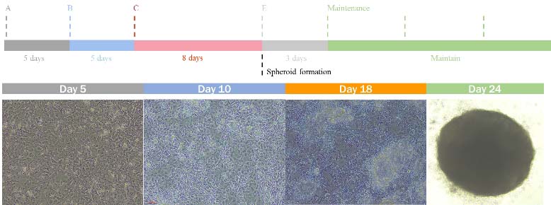
Protocol Diagram of liver organoid differentiation.
Orgnaoid Histology
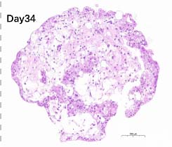
H&E Staining of liver organoid.
Marker Expression
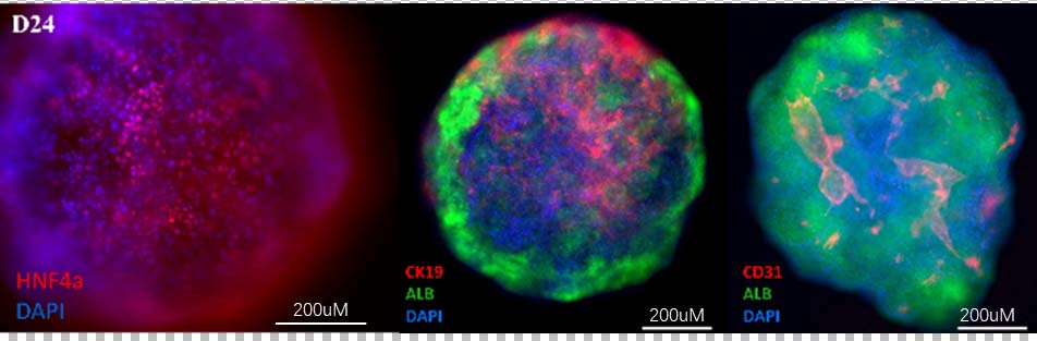
The liver organoids show expression of hepatocytes (HNF4a), hepatobiliary tracts (CK19), Albumin (ALB) and vascular endothelial cell (CD31).
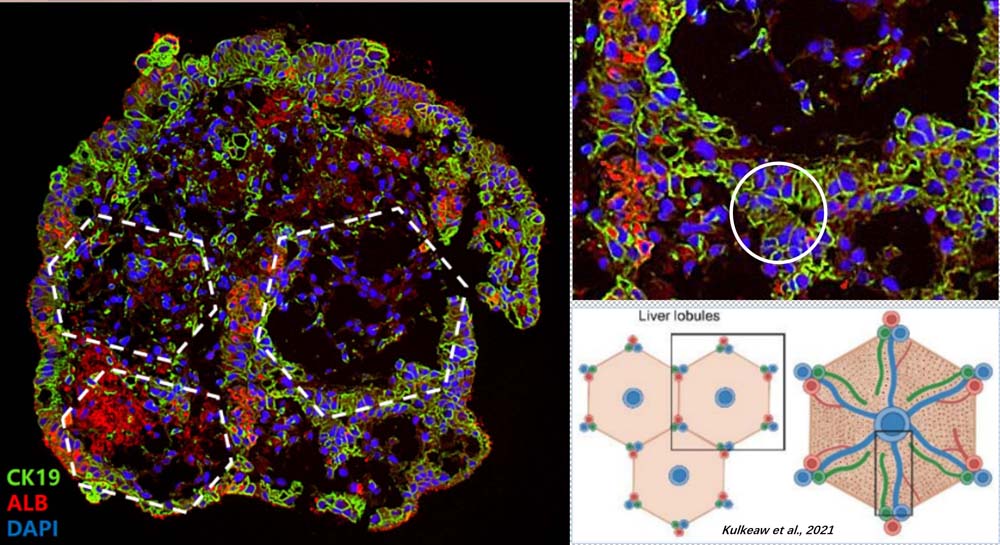
Pre-mature liver organoids show liver lobules-like structure.
Organoid Activity

Liver organoids show better performance than HepG2 cell line in terms of the expression of Albumin, CYP3A4, CYP1A2, CYP2C9, CYP7A1 and BSEP.
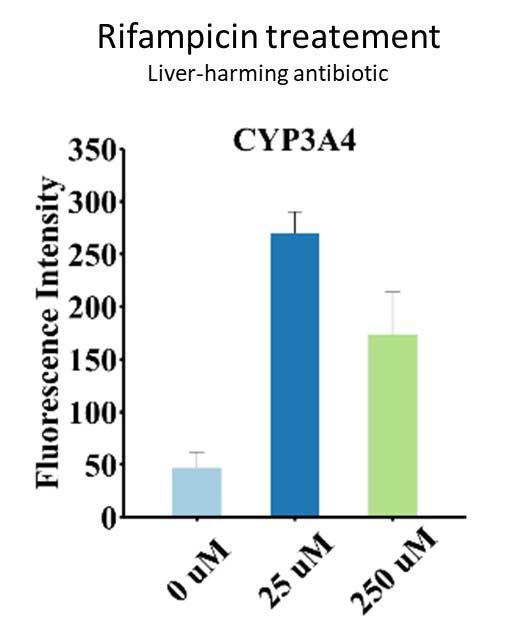
Liver organoid show response to Rifampicin treatment as indication by the increase fluorescence intensity of CYP3A4.

At 20 days of age, the albumin secretion of ACRO's liver organoids was consistent with the secretion amount reported in the literature, being 15 pg Albumin / ng RNA. As the organoids continued to mature and grow, their albumin secretion would keep increasing, reaching a level slightly lower than that of the primary tissue.
Organoid Application

ACRO utilized liver organoids to construct the NASH model. As can be seen from the oil red staining results shown in the above figure, the modeling group of NASH exhibited obvious lipid accumulation (red positive staining).

During the process of constructing the NASH model, we detected the expression of inflammatory factors in liver organoids through qPCR experiments, and observed the upregulation of factors such as IL-6 and IL-33. This confirmed the inflammatory response of our liver organoids.

Liver organoids can be used to detect hepatotoxicity. After treating liver organoids with the commonly hepatotoxic positive drug Trogalitazone, changes in cell viability within the liver organoids can be observed through the results of AO/PI staining.

Fluorescent-free 5-Carboxy-2',7'-Dichlorofluorescein with organoesterase activity are metabolized into fluorescent-emitting CDF, and CDF is actively transported from hepatocytes to the bile duct network and deposited in the bile duct network to simulate the cholestasis process. Trogalitazone treated organoids shows the accumulation of the CDF.
用户评价 发表评论

 联系我们
联系我们 


 项目合作
项目合作